Chapter 2 : The Spinal Cord

Brain: Contents Page

Dorsal Horn Neurones and their Projections

The neurones of the grey matter of the spinal cord are organised into different groupings.

Dorsal Horn neurones receive inputs from afferents entering through the dorsal roots; some of these afferents are myelinated and others are unmyelinated. These different fibre groups have different destinations within the dorsal horn, where the post-synaptic neurones are arranged in layers. Most of the post-synaptic neurones project to the brain, but others have axons that are do not leave the dorsal horn.

Dorsal Horn neurones project to many areas of the forebrain including:

  • The reticular formation of the brainstem
  • The peri-aqueductal grey matter of the midbrain.
  • The thalamus
  • The cerebellum

In addition, dorsal horn neurones receive inputs from various parts of the CNS including the reticular formation and periaqueductal grey matter of the midbrain.

Top

Rexed's Laminae : The Arrangement of Neurones within the Spinal Grey Matter

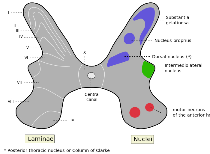
A neuroanatomist called Rexed looked at the structure of the grey matter of the spinal cord and divided it into columns of cells that were similar to each other in different segments.

His observations were based on the shape and size of the neurones, and he divided the grey matter into 10 (Roman X) layers and 6 of these are in the dorsal horn, numbered I to VI .

The classification has been useful in that the different lamina have neurones with different functions.

Neurones involved in Sensation

Lamina I was the marginal layer or zone.

Lamina II had a gelatinous appearance and is sometimes called the substantia gelatinosa.

Lamina IV and V had larger cells and this area has also been called the Nucleus Proprius

In Lamina VI another anatomist gave his name to Clarke's column, sometimes called the nucleus dorsalis.

Lamina X is the area around the central canal

In the ventral horn, lamina VIII and IX contain longitudinal columns of motoneurones.


Afferent Inputs to the Dorsal Horn   Top

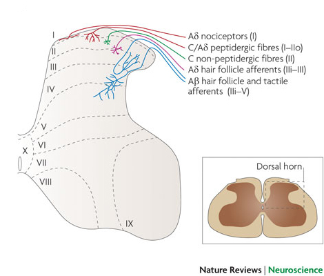
The dorsal horn is concerned with sensory functions, and different types of dorsal root axons originating from the skin and elsewhere synapse in different laminae of the dorsal horn. Some general principles apply: the superficial laminae are concerned with nociceptive sensations, and the deeper laminae also process information from low threshold afferents. However some neurones in the deeper laminae have inputs from nociceptive and tactile receptors : these are called Wide Dynamic Range Neurones (WDR neurones).

Superficial Laminae

Laminae I and II are concerned with processing information from the nociceptive afferents which sense injury and inflammation in their receptive fields and are concerned with pain sensation. The neurones of lamina I, sometimes called marginal cells have long axons that project to the brainstem and thalamus.

Lamina II neurones (the substantia gelatinosa) is concerned with modulating the activity of Lamina I cells. Lamina II cells do not project outside the dorsal horn.


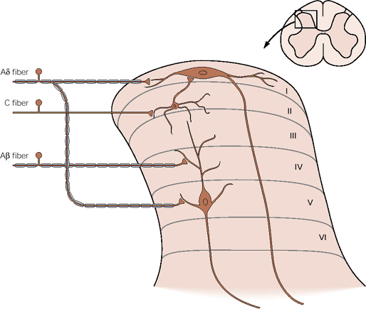
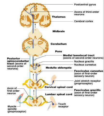
The diagram opposite shows that the neurones of Laminae I and IV/V have axons that project rostrally after crossing the midline of the cords near the central canal.


*

*

Top

Ascending Pathways from the Dorsal Horn

Ascending Pathways from the Dorsal Horn

The unmyelinated and finely myelinated axons of nociceptors and thermoreceptors ('primary afferents') enter the dorsal horn, where they synapse on neurones that project rostrally in the anterior part of the lateral columns on the opposite side of the cord. These neurones collectively constitute the Antero-Lateral System. Some project to the thalamus, hile others project to the brainstem.

Spinothalamic Tract

The neurones of the spino-thalamic tract carry information cross the midline near the central canal, before ascending in the lateral columns and proceeding to the thalamus. The cross-over occurs in the segment of entry and several segments rostral to that. These are sometimes called the 'second-order' neurones.

The diagram opposite shows the spinothalamic tract, and its pathways from the dorsal horn through the lateral brainstem, passed the central grey matter around the aqueduct of the midbrain into the thalamus. There, they synapse on thalamic neurones that project to the cortex and other areas of the CNS.

Spinothalamic Tract

Spino-Reticular Pathway 

The spino-reticular pathway arises in the dorsal horn and ascends in the contralateral white matter to synapse in the reticular formation. Third order neurones in this pathways project to the thalamus, and Fourth order neurones carry the infomation to the cerebral cortex. The destination of these axons include the somatosensory cortex and the association cortex.

Spino-Mesencephalic Pathway

The spino-mesencephalic pathway arises in the dorsal horn and ascends contralaterally to reach the periaqueductal grey matter of the midbrain. This area is involved in modulating nociceptive transmission in the dorsal horn (see below).

Post-Synaptic Dorsal Column Pathway

The classical view of the dorsal columns is that the axons are not concerned with signalling pain. However more modern evidence suggests that, in addition to the axon collaterals of large sensory axons, the dorsal columns also contain post-synaptic axons originating from the dorsal horn.

*

ib.cnea.gov.ar

Spino-cerebellar pathways

Spinocerebellar Tracts

The cerebellum receives an important input from muscle and joint receptors that carry information about the position of the limbs.

Each side of the cerebellum is concerned with coordination of muscles on the SAME side of the body - hence the spinocerebellar tracts are an ipsilateral rostral projection from the cord (unlike the anterolateral pathways).

The diagram opposite shows, on the left hand side of the spinal cord, the pathway that carries proprioceptive information to the cerebellum. It involves afferent inputs from muscle and joint receptors that end in a nucleus at the base of the ipsilateral dorsal horn; this is Clarke's column (or the nucleus dorsalis). Axons arising from cells in that nucleus proceed to the ispilateral cerebellum.

More details of the spino-cerebellar system can be found in the following link. Ascending Pathways to the Cerebellum


The Dorsal Column-Medial Lemniscal System is dealt with in greater detail in the next Chapter, and carries infomation about Fine Touch, Vibration and Proprioception to the Cerebral Cortex.

The Spino-Cerebellar pathway and the

Dorsal Column-Medial Lemniscal System

*


Todd, Nature Neuroscience Reviews
http://www.ib.cnea.gov.ar/

Top
Chapter 2 : The Spinal Cord

Brain: Contents Page